
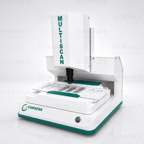
Multiscan - Digitales und automatisiertes Mikroskopiesystem
Beschreibung
Why Multiscan?
- Reduces staff workload through automated scanning
- Image acquisition with reproducible settings
- Maintain existing workflows: continue using familiar sample preparation methods and sample carriers
- Traceable digital documentation
- Optional: AI support for selected applications
The Multiscan is an automated microscopy system for the digital acquisition and documentation of microscopic samples. The system was developed for routine applications requiring standardized image acquisition, reproducible results and digital processing of the acquired data.
The Difference Between Basic and Pro
The main difference between the Basic and Pro models lies in the scanning area and, consequently, the sample throughput. The Basic model can automatically scan up to 3 slides per run. The Pro version features an additional axis, providing a larger scanning area and allowing up to 8 slides to be automatically scanned in a single run.
The Workflow
Sample acquisition is performed automatically via a motorized scanning process. An autofocus function supports reproducible image acquisition. The acquired image data is immediately available for further evaluation, documentation or archiving.
From Sample to Documented Result
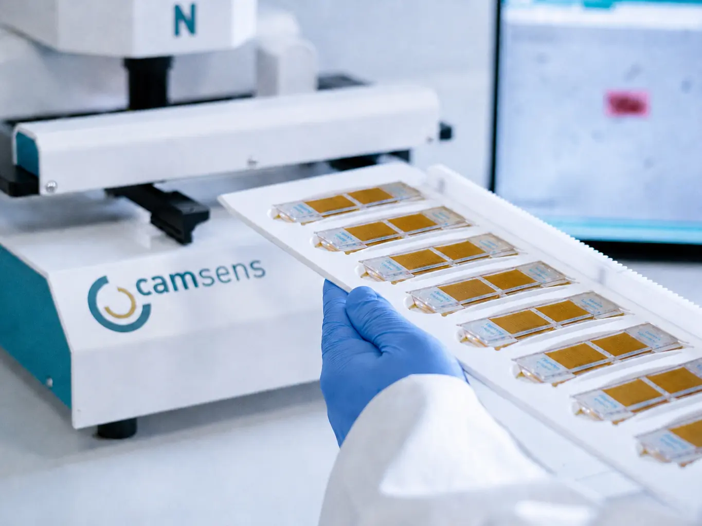
Prepare & Load
Prepare one or more samples using established methods and load them into the Multiscan.
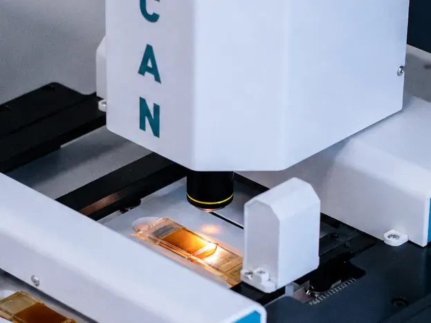
Automatic Scanning
Multiscan automatically scans defined areas using autofocus, Z-stacking and reproducible settings.
Review, Analyse & Document
Review digital results, use AI models if required, and document findings for traceable reports and easy sharing.
One System. Multiple Sample Formats.
The Multiscan supports different sample carrier formats and can be configured for various microscopy applications. Depending on the requirements, different objectives and scanning parameters are available, allowing the system to be adapted to the respective examination task. These include conventional microscope slides as well as other sample carriers used in routine analysis.
Sample Formats:
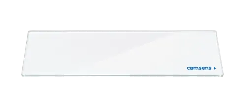
Microscope Slides (Image) (up to 3 (Basic),
up to 8 (Pro) per run)
Automated digital scanning for efficient evaluation, documentation and sharing.
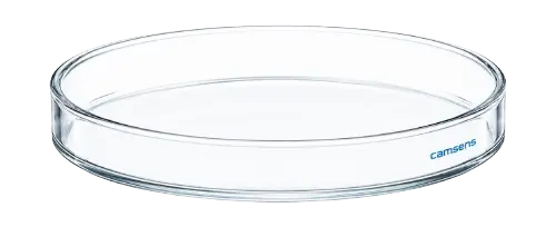
Petri Dishes (Image) (up to 3 per run)
Scanning of Petri dishes of different sizes for microbiological and other imaging workflows.
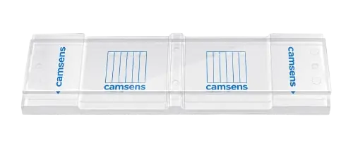
McMaster Counting Chambers (Image) (up to 3 (Basic),
up to 8 (Pro) per run)
Digitization of established counting procedures for standardized counting and evaluation.
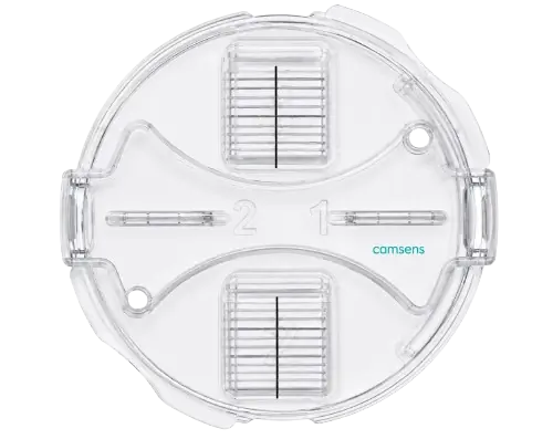
Mini-FLOTAC (Image) (up to 3 per run)
Digitization of Mini-FLOTAC preparations for parasitological evaluation and documentation.
Standardized image acquisition helps reduce variations that may arise from manual operation. Automated sample processing supports consistent data acquisition and enables multiple samples to be processed according to defined parameters. This makes the system particularly suitable for laboratory environments with regularly recurring examinations and an increased need for documented workflows.
The Multiscan can be used in research, diagnostics, quality control and other microscopic laboratory applications.
Application Areas
The device is flexible and adaptable. It can be used in various fields where biological samples are examined.
Veterinary Medicine, Biomedical Research, Life Science
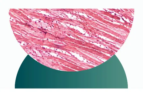
Digital Scanning of Microscope Slides
Automate the digital scanning of microscope slides with configurable scanning areas, Z-stacks and objectives for digital evaluation and documentation.
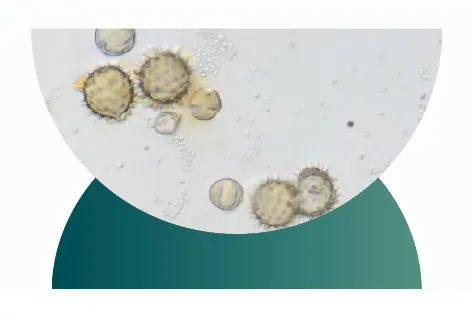
Application Development
Develop application-specific microscopy workflows with standardized image acquisition, digital documentation and customized AI models.
Microbiology
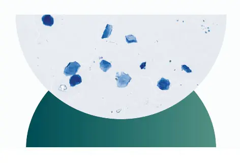
Microbiological Air Analysis
Automate image acquisition of samples containing airborne spores and particles, then review, analyse and digitally document the results at the workstation.
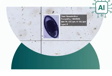
AI Applications – Pre-trained Model: Veterinary Medicine – Parasitology
AI-assisted counting of parasite eggs and oocysts in ruminants, horses, dogs and cats (ready-to-use models). Multiscan automates routine sample analysis for parasite detection and delivers results within just a few minutes.
Technical Features
- Automated digital image acquisition of microscopic samples
- Motorized scanning of defined sample areas
- Integrated autofocus function
- Support for various sample carrier formats
- Digital documentation and archiving of image data
- Customizable scanning parameters
- Software-assisted evaluation and data management
- Standardized and traceable workflows
- 4×, 10×, 20×, 40× and 60× objectives
- Compact dimensions: W 30 × D 40 × H 44 cm, 10 kg
- 5 MP Camera
Weitere Informationen
| Abmessung | 30×40× 44cm (BxTxH) |
|---|---|
| Verpackungseinheit | 1 Stück |
| Gewicht | 10kg |