Ein Farbstoff für FIRM – Fluorophore-Infiltrated Resin Microscopy
Beschreibung
Funktionsprinzip:
Das Prinzip dieser Methode beruht auf der Infiltration dünner in Harz eingebetteter Schnitte mit einem speziellen Fluorophor.
Bei Betrachtung mittels Fluoreszenzmikroskop, leuchtet das Harz hell, Zellen und Gewebestrukturen hingegen bleiben dunkel in einem negativen Relief.
- Verwendbar mit Epoxid- und Acrylharzen
- Einzigartige Färbemethode: der Fluoreszenzfarbstoff dringt in das Harz ein ohne Anfärbung der Gewebestrukturen (negatives Relief)
- Sehr hoher Kontrast und Auflösung
- Prüfung der Proben vor der Abbildung im EM
- Ideal auch für analytische Mikroskopie in Material- und Lebensmittelforschung
Protokoll zur Anwendung:
- Laden von Semidünnschnitten auf Glasobjektträger
- An Luft antrocknen und Zugabe von 100µl FIRM Farbstoff für 30 Sekunden
- Abspülen und mit Wasser oder einem wässrigen Medium Eindecken
- Aufnahme im Rhodamin-Kanal eines Standard-Fluoreszenzmikroskops
Human anterior pituitary, FIRM, rhodamine channel. Note that the fluorophore illuminates the LR White resin, providing an image of tissue structure primarily in negative relief. Bar, 100µm. All subsequent FIRM images are shown in monochrome.

Human anterior pituitary, formalin-fixed autopsy specimen, comparison of H&E paraffin section (left) with FIRM (right). 20x dry objective used for both. Scale bar, 20µm.

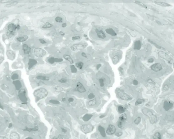
Human anterior pituitary, FIRM image taken with 150X glycerin objective. Individual secretory granules are easily visualized, ranging from 150- 350nm in diameter. Scale bar, 5µm.

Correlative FIRM/fluorescent lectin labeling of human pituitary. A single LR White thin section was labeled first with Concanavalin AAlexa-488 (B) imaged, then stained for FIRM imaging (A). Densely granulated cells (arrows in A) are strongly labeled with Con-A (arrows in B). Dark structures resembling large lysosomes or residual bodies (arrowheads in A) are also strongly labeled by Con-A, consistent with reports in the literature.

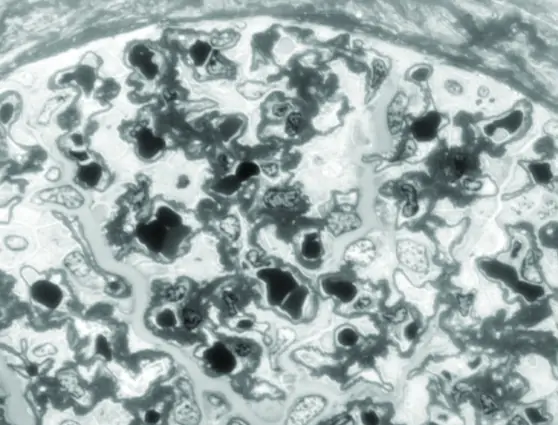
Comparison of toluidine blue staining (top) to FIRM (middle) on near adjacent sections of human kidney. Note enhanced contrast, especially of glomerular basement membranes in FIRM image. High power FIRM image (bottom) of glomerulus. Note resolution of podocyte foot processes (arrows). Scale bars in A, B, 20µm, in C, 5µm.